
In the field of clinical microbiology, there is a need for a deeper understanding of the intricate relationship between bacteria and eukaryotic cells, particularly in terms of the microbiome and the immune system’s ability to identify and respond to infection. This aspect of the relationship is not given sufficient emphasis at the bench. However, it could be argued that grounding in this understanding can facilitate the bench practitioner to apply an extra layer of understanding that Standard operating procedures and algorithms cannot teach.
To grasp the complexity of this relationship, knowledge in both microbiology and immunology is required, along with an understanding of the functions of the innate and adaptive immune responses.
The innate and adaptive immune responses are interconnected components of the immune system that collaborate to defend the body against pathogens and maintain immune homeostasis. The innate immune response is non-specific and rapid, and is observed in most, if not all, eukaryotes. It primarily consists of physical and chemical barriers, such as the skin and mucus membranes, as well as various proteins and enzymes that provide chemical defense. Additionally, cells of the innate immunity, such as macrophages and dendritic cells, possess pattern recognition receptors that can detect changes in the internal environment by scanning for self and non-self antigens.. These cells produce pro-inflammatory mediators, which promote the recruitment of immune cells, such as neutrophils and macrophages, to the site of infection, enabling the engulfment of pathogens through phagocytosis. Furthermore, other types of cell such as natural killer cells are capable of identifying the absence of MHC class I, allowing them to detect cancerous cells, or viral antigens on their peptide scaffold. For ease of conversation, there are two classes of major histocompatibility complex (MHC). Class, one deals with primarily endogenous proteins and proteins from viral antigens, and class two MHC expresses exogenous proteins as a byproduct of proteolysis within phagocytic cells.
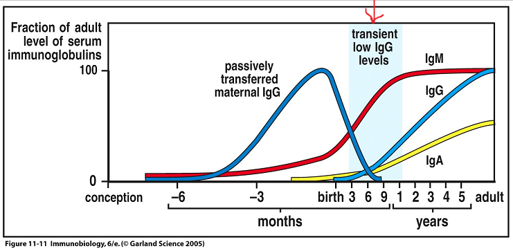
In contrast to the innate immune response, the adaptive immune system is slower and highly specific. It takes time to develop, but it provides targeted defense against antigens. This is exemplified in infancy, where newborns receive antibody protection from their mothers and subsequently develop their own immune competence over time.
The adaptive immune response relies on the recognition of specific antigens by T cells and B cells. The process involves interaction between the antigen and antigen-presenting cells, such as macrophages, which initiate clonal expansion and eventually lead to antibody production as B cells develop into plasma fells and eventually memory cells. Communication between the external and internal environments of the antigen-presenting cell occurs through intricate pathways involving phosphorylation and activation of the ZAP-70 pathway via toll-like receptors.
Toll-like receptors (TLRs) are a family of proteins located on the surface of immune cells. When a pathogen invades our body (such as bacteria, virus or protozoan), TLRs recognize specific components of the pathogen (foreign antigen) and initiate the immune response. Each TLR is specialized in recognizing specific molecular patterns (PAMPS) associated with different types of pathogens. This recognition triggers a signaling cascade within the immune cell. This cascade activates various immune responses, such as the production of inflammatory cytokines, chemokines, and antimicrobial peptides, to fight off the infection.
Stop and think: reflect on areas of your clinical practice, where you have encountered a patient with a potentially infected wound, or even potentially sepsis. What are the clinical signs of infection you have observed and how do they correlate with the immune response above? 👆
TLRs not only play a direct role in immune defense but also help to shape the adaptive immune response. By activating immune cells and releasing signaling molecules, TLRs promote the activation and maturation of other immune cells, such as T cells and B cells.
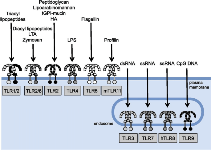
The image below depicts TLR’s associated with respective antigens (Ligands)
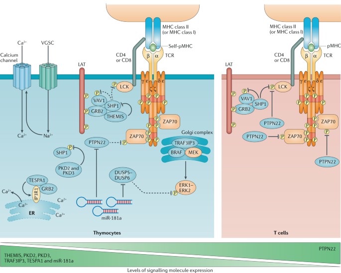
The ZAP-70 pathway is crucial in the activation of T cells, which coordinate the immune response against pathogens and abnormal cells. T cell receptor activation triggers a series of signaling events, beginning with the recruitment and activation of CD3 and ZAP-70. CD3 activation occurs through phosphorylation, creating docking sites for ZAP-70, an important molecule in cell signaling. ZAP-70 itself is phosphorylated by lymphocyte-specific protein tyrosine kinase, leading to the phosphorylation of the scaffolding protein LAT (link for activation of T cells). LAT is implicated in the recruitment of other T cells. The downstream signaling process is complex, ultimately resulting in the release of calcium and activation of protein kinase C from the endoplasmic reticulum. Phosphorylation activates transcription factors that regulate gene expression, modulating T cell activation and function.
Understanding of the disease sequelae before specimens arrive at the microbiology lab is an important feature when considering clinical information provided. Immunology and microbiology combined provide a cornerstone for infection control and prevention strategies in addition to disease management and provide useful epidemiological insights.
Gaud, G., Lesourne, R. & Love, P.E. Regulatory mechanisms in T cell receptor signalling. Nat Rev Immunol 18, 485–497 (2018). https://doi.org/10.1038/s41577-018-0020-8

 The random ramblings of an aspiring BMS